Published On Jun 19, 2024
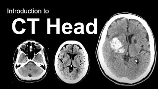
Video on the basis of CT brain, aimed at medical students and radiology residents at the start of their training. Everything you need to know that cover that first night shift at the ER: how a CT image is created, basic interpretation of CT images, brain anatomy and how to recognize acute ischmemic stroke, intracranial hemorrhage, brain herniation and hydrocephalus.
0:00 - Introduction
1:15 - Basic principles of CT
7:52 - Density and the Hounsfield scale
13:34 - Windowing your images
16:28 - Brain window and bone window
19:02 - Stroke window
20:51 - Subdural window
22:49 - CT artifacts
24:00 - Beam hardening artifacts
28:41 - Brain Anatomy on CT
29:27 - The skull
32:04 - The Meninges
36:16 - The CSF-spaces: sulci, fissures, ventricles and cisterns
43:57 - The cerebral cortex
45:26 - The deep nuclei
46:42 - The internal capsule, corona radiata and centrum semi-ovale
48:48 - The corpus callosum
49:42 - The posterior fossa
50:19 - Brain Pathology on CT
51:30 - Quick CT check for pathology
53:27 - Acute ischemic stroke
1:01:33 - Brain hemorrhage
1:12:52 - Brain herniation
1:18:29 - Hydrocephalus
1:23:54 - Herpes encephalitis, diffuse brain edema, PRES
1:27:21 - Key Messages
This video is brought to you by the neuroradiologist:
/ theneuroradguy
/ the_neurora. .
https://theneuroradiologist.org/
#radiology #neuroradiology #neurology #medicalstudent #neuroradiologist #theneuroradiologist #MRI #CT #medical #pediatrics #mri #radiologytechnologist #radiologyresident #brainanatomy