Published On Nov 14, 2020
Hello everyone!!
Thank you very much for your appreciation of our EDUSURG channel in the "small talk series" as well as the "step by step surgery series" that started with laparoscopic cholecystectomy.
Now, we get back to our liver anatomy STS series and finish the Basic liver anatomy series with the liver imaging and identification of liver segments on MRI. We will be discussing some advanced liver anatomy concepts such as liver arterial variations and biliary variations in a separate video after the basic series is over so STAY TUNED for that...
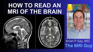
In the current video, liver anatomy STS now enters its last phase where we discuss MRI liver. MRI of liver is one of the most tricky MRI scans to study and hence, it is discussed here from MRI basics to liver MRI basics and then liver segmental anatomy on MRI
A KEY POINT TO HIGHLIGHT AGAIN is "FOR ANY DOCTOR WHO WISHES TO MANAGE LIVER DISEASE PATIENTS, LEARN TO SEE THE SCANS FOR YOURSELF AND WITH YOUR RADIOLOGY COLLEAGUES ON THE CONSOLE"
This is true for all diseases but, especially for liver and pancreas surgeries where a multidisciplinary discussion of the case with all is of paramount importance.
This video is, therefore, very important because we need to identify the liver segments on an MRI scan as only then, can we identify lesions and plan surgeries on the liver.
BASIC MRI SEQUENCES ARE DESCRIBED IN THE VIDEO FIRST IN BRIEF
T1 non-fat suppressed or non-fat saturated
T2
T1 fat-suppressed or fat saturated
Heavily T2 weighted or MRCP
Contrast MRI (T1 fat suppressed images timed at different phases)
After this, we dive into the couinaud classification on liver MRI sequences. Liver MRI is also called a TRIPHASIC scan LIVER PROTOCOL with
Arterial phase or a late arterial phase
Portal venous phase and
Delayed or hepatic venous phase
If we use a hepatobiliary specific contrast, you can also have a hepatic phase at 15-20 minutes, but, this we will discuss in advanced MRI sections later.
In this video, We have also highlighted a few space-occupying lesions in the liver to show you how the concepts in the video can help you localize the segment of the liver space-occupying lesion.
KINDLY MIND THE MINOR VOICE LAG IN THE VIDEO WITH REGARDS TO THE CURSOR MOVEMENT.
WE HAVE CHANGED THE SOFTWARE NOW AND THIS POINT WILL BE TAKEN CARE OF IN THE SUBSEQUENT VIDEOS ON RADIOLOGY.
This video series will help you understand the basics of liver anatomy and will help you in
Writing surgery questions in the exam
Answering the questions in practical exams
Multiple choice questions on surgical anatomy
Understanding and planning liver surgeries on the scan.
So. let's have fun...
Do SHARE, SUBSCRIBE, LIKE if you like the approach to this video.
Also, LEAVE TOPICS, SUGGESTIONS in comments.
Let's make surgery fun, factual, and free...